SPECT measures how well the brain is working by scanning blood flow in the brain. Injured areas will have less blood flow. It allows the specialist to see a picture of your brain without opening up your skull. It takes a skilled radiologist to be able to read the 3D images and 2D slices, though.
“Imaging cerebral blood flow is used to assess brain function. These studies use several SPECT radiopharmaceuticals and mostly are technetium-99m imaging agents. They include technetium-99m pertechnetate, technetium-99m-pentetate (Tc-DTPA), technetium-99m gluceptate (Tc-GH), technetium-99m exametazime (Tc-HMPAO) and technetium-99m bicisate (Tc-ECD).
All gamma radiation emitted from SPECT radiopharmaceuticals are detected by the usage of a rotating gamma camera around the patient to produce 3-D imaging. The images undergo various electronic transformations by taking into account the distribution of the radiotracer, the process of filtered back projection and other tomographic techniques.
The radioisotopes used in SPECT scanning have relatively long half-lives, for example, technetium-99m (t½ = 6 hours)”
SPECT imaging applications to myocardial perfusion and brain imaging
Half-life is the amount of time for half of a radioactive substance to degrade and lose its radioactivity. It’s also used to describe how long a drug remains effective. So in this case, half of the technetium-99m becomes non-radioactive in 6 hours. The half that’s left becomes non-radioactive in the next 6 hours, and so on. Drinking lots of water or your non-alcoholic drink of choice after the scan will help you rid your body of the tracer. Wash your hands well after using the bathroom. In the pandemic age, this should be a habit, anyway, many times during the day. In about 24 hours, what radioactivity is left won’t be much.
The How of SPECT
The SPECT scanner reads gamma rays emitted from a radioactive tracer injected in your arm about one hour before the scan. Which tracer depends on what is being scanned.
You’ll be sent to wait in a waiting room while the tracer makes its way to your brain. When it’s time to scan you, the technician will come and get you and have you lie down on a narrow bed with your head supported. You must keep your head still during the scan. Some places will distract you; some will be utilitarian and you have to keep your own mind engaged. It’s much harder to stay still when you’re not distracted with music or a show. It may be helpful to have someone accompany you to keep your mind occupied.
Your test location ought to have an online explanation, but I’ve discovered how informative that can be varies greatly. For example, UHN, a hospital network in Toronto, versus HealthLink BC in British Columbia: the former almost reads like they’re talking to a child, the latter is fairly comprehensive.
This video shows what it’s like, although I only had to wait about an hour after the injection of radioactive tracer before I was scanned.
Resources
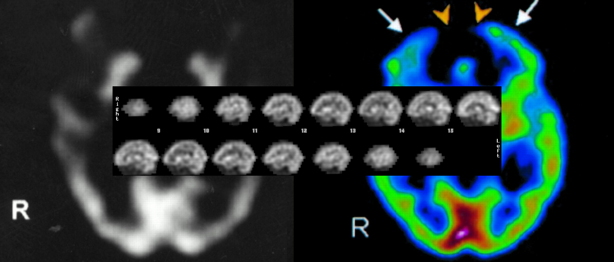
Large SPECT images at top from Ana M. Catafau. Brain SPECT in Clinical Practice. Part I: Perfusion. Journal of Nuclear Medicine. Vol. 42, Issue 2. pp 259-271. 1 February 2001