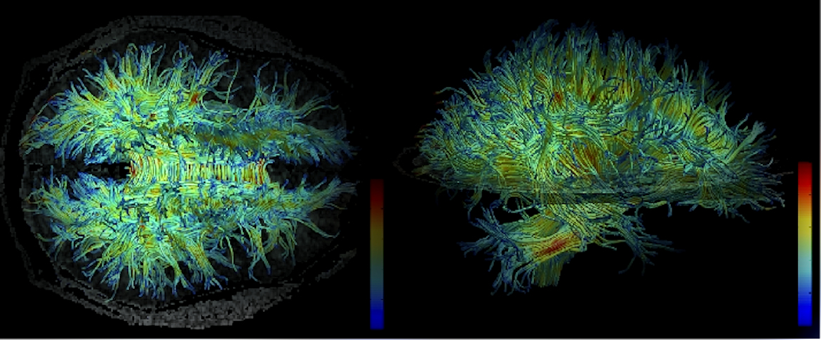
Diffusion tensor imaging is a type of MRI that maps white matter tractography. Tractography visually represents the bundles of neurons that have myelin sheathes. Myelin sheathes make the neurons look “white” and increase the speed of transmission of electrons down the neuron. DTI is used to study and treat neurological disorders. It apparently reveals the extent of injury to the networks of white matter, that is, brain networks. Having this kind of imaging helps to understand one’s difficulties and how to treat them. Like MRI, it’s non-invasive and doesn’t necessarily require tracer to be injected.
“DTI technique was first introduced by Peter Basser in 1994. It is an improved version of conventional MRI wherein signals are solely generated from the movement of water molecules. The term ‘diffusion’ denotes random thermal motion of water molecules. In other words, DTI uses the diffusion of water as a probe to determine the anatomy of a brain network, which basically provides information on static anatomy that is not influenced by brain functions.”
Dr. Sanchari Sinha Dutta, Ph.D. Diffusion Tensor Imaging (DTI) Explained. News Medical Life Sciences.
What does this mean? Water will travel faster along the length of an axon — the route of electrical conduction down a neuron or nerve cell — than perpendicular to its length. In addition, whether a neuron is myelinated or not determines how quickly water will travel along the axon. Myelin increases speed of electrical conduction, and also water movement. This provides a picture of axonal direction, neuronal myelination, and the orientation of nerve fiber tracts. That means DTI will pick up on injury that damages neurons or demyelinates them and injury to neural networks, that is, nerve fiber tracts.
The How of DTI
DTI is like getting an MRI. You’ll change into a hospital gown and pants and remove all metal. You may need lotion to help you remove rings. Because this scanner uses a large magnet, and magnets attract most metal, you don’t want anything metal to be ripped from your body.
You’ll be taken to a large room where the enormous scanner awaits. You’ll lie down on a narrow bed that slides into the machine. Newer machines are more open to reduce the effect of claustrophobia. The older machines are claustrophobic because you are fully inside basically a narrow tunnel. Some hospitals will provide you with a washcloth to cover your eyes (yes, it does help with claustrophobia not to see how close the scanner is) and music, either through headphones or the machine. Others will provide no distraction. All should provide you with earplugs. The scanner is loud. As in deafening. It’s not a hum. It’s a series of rapid and slower clicks and bangs. You must remain completely still during the scan.
Some clinics will provide sedatives to help you cope with claustrophobia. A word of warning to those with brain injury: the sedative may do nothing for your anxiety because brain injury changes the way drugs work. Even if you have taken the same one before brain injury, don’t expect it to work in the familiar way after. For me, I became as if I was drunk with zero change in my claustrophobia. You can ascertain from the clinic or hospital beforehand how long the scan will be. Be prepared to have it scheduled at any time of day or night.